
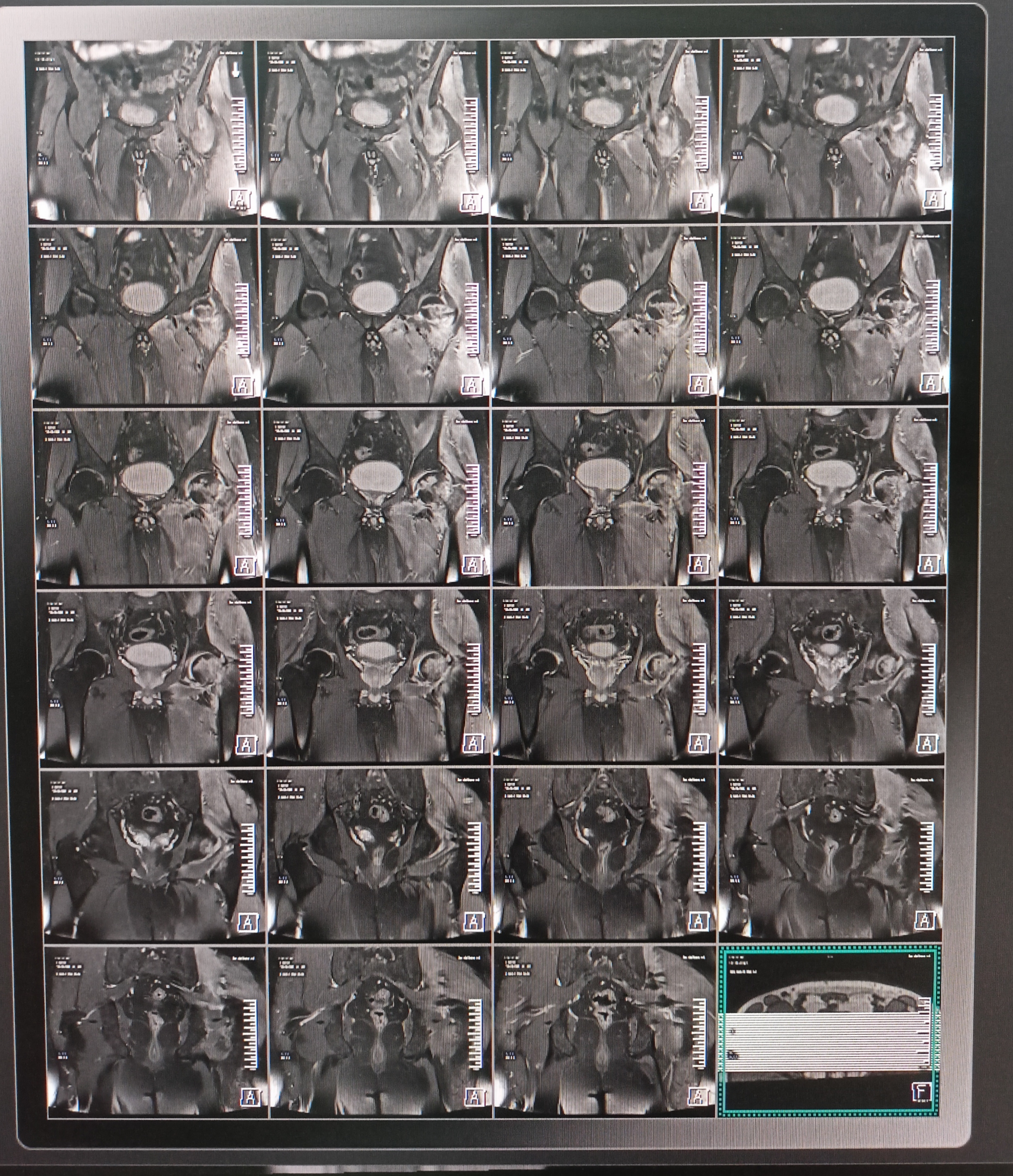
mri hip joint
A hip MRI scan is an imaging exam that examines the pelvic bones and hip joints. The hip joint, where the pelvis is attached to the legs, is a ball and socket synovial joint. It connects the pelvic acetabulum and the top of the femur and allows movement of the mid-body
MRI Hip joints
1.FS coronal
2.t1 coronal
3.Pd fs transverse
4.t1 transverse
5.t2 transverse
6.Fs sagittal RT hip
7.Fs sagittal LT hip
FS coronal 
t1 coronal 
Pd fs transverse 
t1 transverse 
t2 transverse 
Fs sagittal RT hip 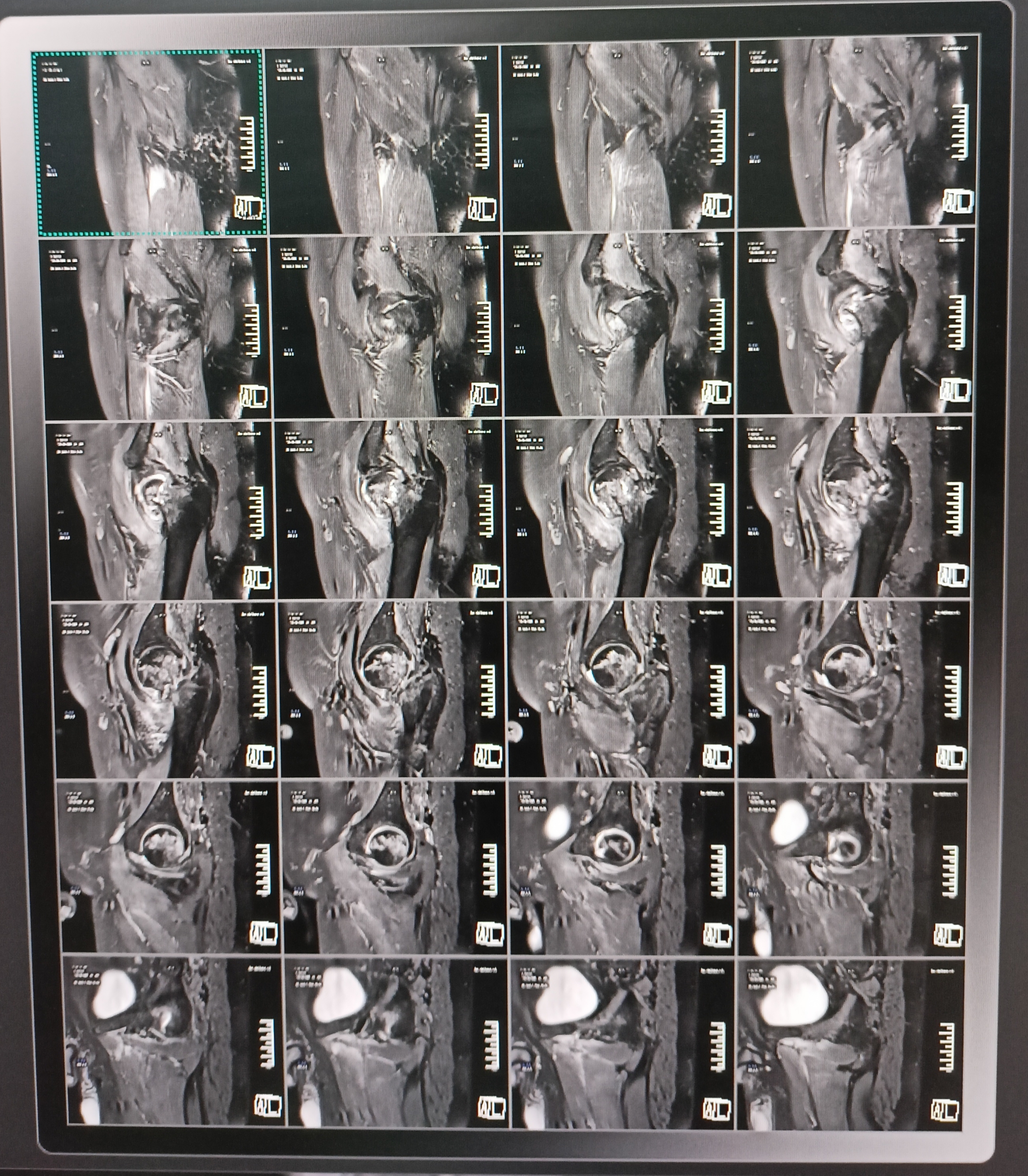
Fs sagittal LT hip
No comments