
mri pituitary with contrast
A systematic approach to the pituitary region is crucial as small lesions can have a profound impact on the patient, and can be subtle even on high-quality dedicated MRI imaging.
Successful assessment of the pituitary region relies not only on a clear understanding of the local anatomy but also on the relatively wide variety of pathologies that occur in the region.
MRI Film
1.Contrast t1 coronal & sagittal 2.5mm
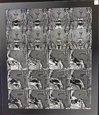
2.Contrast dynamic 1st,2nd,3th,4th,5th images
3.Contrast dynamic 1st,2nd,3th,4th,5th images
4.NC t2 coronal & t1 coronal 2.5 mm
5.NC t1 sagittal 2.5 mm
6.DWI+ADC
7.t1 sagittal
8.t2 coronal
9.t1 transverse or axial
10.t2 flair transverse or axial
11.t2 transverse or axial
 |
| Contrast t1 coronal & sagittal 2.5mm |
 |
| Contrast dynamic 1st,2nd,3th,4th,5th images |
 |
| Contrast dynamic 1st,2nd,3th,4th,5th images |
 |
| NC t2 coronal & t1 coronal 2.5 mm |
 |
| NC t1 sagittal 2.5 mm |
 |
| DWI+ADC |
 |
| t1 sagittal |
 |
| t2 coronal |
 |
| t1 transverse or axial |
 |
| t2 flair transverse or axial |
 |
| t2 transverse or axial |
Thanks for visit
No comments